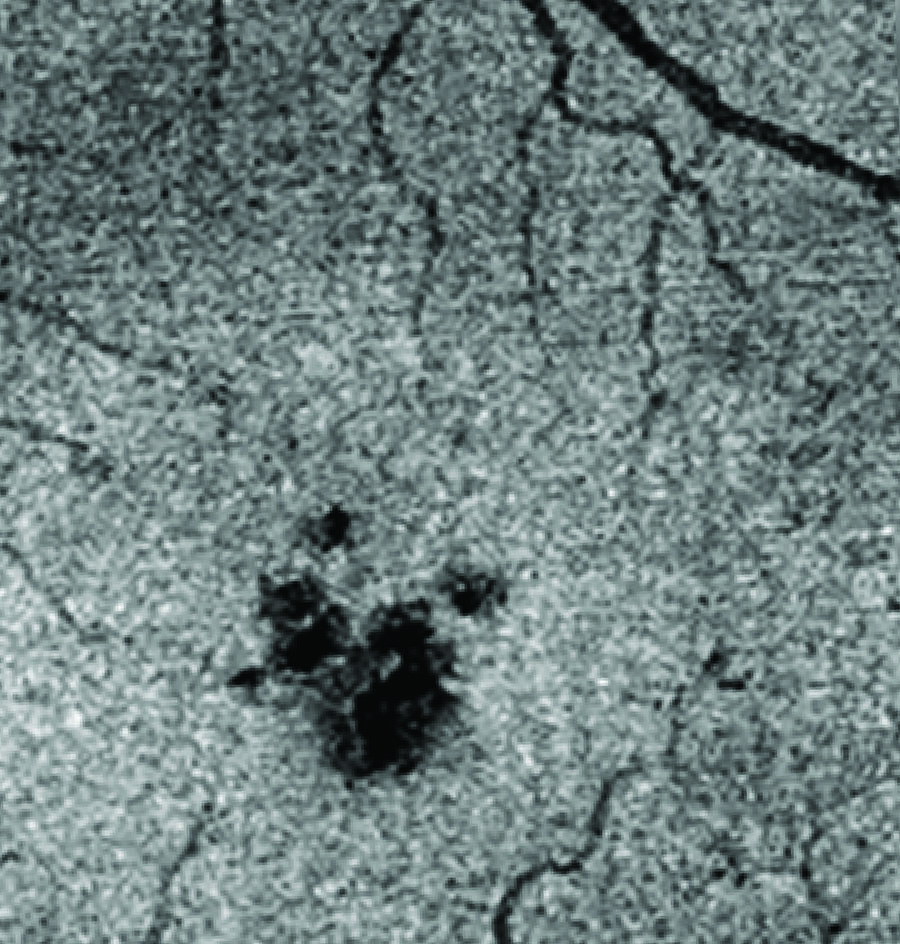
This en face OCT image of a MacTel eye shows a disruption in photoreceptors (dark patch) in the foveal region of the retina.
Advanced imaging technologies have changed our understanding of disease progression in macular telangiectasia type 2. Clinicians affiliated with the Lowy Medical Research Institute are using new imaging modalities to diagnose and follow changes in the MacTel eye. Some of these imaging modalities were developed for research purposes, or are in early stages of development with manufacturers. Many such specialized devices are not yet widely available across all LMRI-affiliated clinical sites.
Some of these technologies capture very high-resolution pictures of the retina, in some cases down to the cellular level. For example, scientists can see individual photoreceptors by adaptive optics imaging. Some clinicians who participate in the MacTel Project have specialized OCT devices that allow them to see blood vessels in the eye, with optical coherence tomography-angiography (OCT-A) machines. Recent research indicates that the MacTel retina has a signature pattern observed by fluorescence lifetime imaging ophthalmoscopy (FLIO), which may allow for earlier diagnosis in the future, or diagnosis among family members. While not widespread, such specialized imaging devices are very important to MacTel research.
Other imaging technologies, including optical coherence tomography (OCT) and microperimetry, are in use by members of the MacTel clinical community around the world.
Diagnosing Mactel with Retinal Imaging
Fluorescein angiography is commonly used to detect vascular alterations that occur due to MacTel. Fluorescein angiography typically shows telangiectatic capillaries temporal to the foveola in early-stage MacTel. In late-stage disease, fluorescein angiography shows a diffuse hyperfluorescence. It should be noted that MacTel causes retinal changes that cannot be seen by fluorescein angiography. This tool is not be effective, alone, to detect early cases of MacTel.
Optical coherence tomography (OCT) is a non-invasive imaging technique that uses long-wavelength light to generate high-resolution images of the retina in cross-section. OCT can be used to detect changes related to MacTel at very early stages. OCT shows asymmetry in the foveal dip very early. It also shows hyporeflective cavities of the inner and outer neurosensory retina, and increased reflectivity of inner neurosensory layers. Importantly, OCT is used to measure the IS/OS break, which is the disruption of the border between inner and outer photoreceptor segments, and has been one of the inclusion criteria for participation in clinical trials.
Confocal Reflectance Imaging with a blue laser at 488 nm shows an increased reflectance in the parafoveolar region of MacTel eyes, surrounded by decreased reflectivity. This has also been referred to as “blue light reflectance imaging.” The images can be captured with a fundus camera and confocal scanning laser ophthalmoscope. The area of increased reflectance is coincident with the loss of macular pigment. Imaging macular pigment loss is a more reliable marker of early disease.
Macular Pigment Density can be imaged in MacTel by a variety of techniques: two-wavelength fundus autofluorescence, two-wavelength reflectance maps, and spectral fundus reflectometry.
Confocal Adaptive Optics Scanning Laser Ophthalmoscopy is a non-invasive imaging technique that can be used to see individual photoreceptors in the back of the eye, along with other features, like cysts, blood vessels, and the RPE cell layer. Using adaptive optics, it is possible to begin to understand how MacTel affects photoreceptor health and function. Adaptive optics is time-consuming and requires very specialized instrumentation. Adaptive optics imaging is not widely available, and is not appropriate for diagnostic purposes, but it is being used in the ongoing clinical trials.
Belfast Ophthalmic Reading Center
An ophthalmology reading center is a specialized facility that evaluates and grades retinal images for clinical studies. While the images may be collected from around the world, centralizing their evaluation ensures data consistency. LMRI works with a longstanding team of expert graders who evaluate registry images for diagnostic features of MacTel. Our designated reading center is the Queen’s University Belfast Ophthalmic Image Reading Center, located in Belfast Northern Ireland. We also work with grading and clinical experts at Moorfields Eye Hospital in London, UK who also grade and diagnose MacTel images.
Using a centralized reading center for all collected images is imperative to our research initiatives as the Reading Center provides an unbiased, third-party analysis of the data. Having a team of experienced graders trained in diagnosing MacTel is especially important for our research, as the disease can be challenging to diagnose and clinically characterize.